Physicists smash particle imitators
Physicists of all stripes seem to have one thing in common: They love smashing things together. This time-honored tradition has now been expanded from familiar particles like electrons, protons, and atomic nuclei to quasiparticles, which act like particles, but aren’t.

Quasiparticles are formed from groups of particles in a solid material that collectively behave like a unified particle (SN: 10/18/14, p. 22). The first quasiparticle collider, described May 11 in Nature, allows scientists to probe the faux-particles’ behavior. It’s a tool that could potentially lead researchers to improved materials for solar cells and electronics applications.
“Colliding particles is really something that has taught us so much,” says physicist Peter Hommelhoff of the University of Erlangen-Nuremberg in Germany, who was not involved with the research. Colliding quasiparticles “is really interesting and it’s really new and pretty fantastic.”

It’s a challenge to control these fleeting faux-particles. “They are very short-lived and you cannot take them out of their natural habitat,” says physicist Rupert Huber of University of Regensburg in Germany, a coauthor of the study. But quasiparticles are a useful way for physicists to understand how large numbers of particles interact in a solid.

One quasiparticle, known as a hole, results from a missing electron that produces a void in a sea of electrons. The hole moves around the material, behaving like a positively charged particle. Its apparent movement is the result of many jostling electrons.

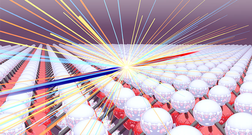
The new quasiparticle collider works by slamming holes into electrons. Using a short pulse of light, the researchers created pairs of electrons and holes in a material called tungsten diselenide. Then, using an infrared pulse of light to produce an oscillating electric field, the researchers ripped the electrons and holes apart and slammed them back together again at speeds of thousands of kilometers per second — all within about 10 millionths of a billionth of a second.

The smashup left its imprint in light emitted in the aftermath, which researchers analyzed to study the properties of the collision. For example, when holes get together with electrons, they can bind into an atomlike state known as an exciton. The researchers used their collider to estimate the excitons’ binding energy — a measure of the effort required to separate the pair.
The collider could be useful for understanding how quasiparticles behave in materials — how they move, interact and collide. Such quasiparticle properties are particularly pertinent for materials used in solar cells, Huber says. When sunlight is absorbed in solar cells, it produces pairs of electrons and holes that must be separated and harvested to produce electricity.

The researchers also hope to study quasiparticles in other materials, like graphene, a sheet of carbon one atom thick (SN: 08/13/11, p. 26). Scientists hope to use graphene to create superthin, flexible electronics, among other applications. Graphene has a wealth of unusual properties, not least of which is that its electrons can be thought of as quasiparticles; unlike typical electrons, they behave like they are massless.
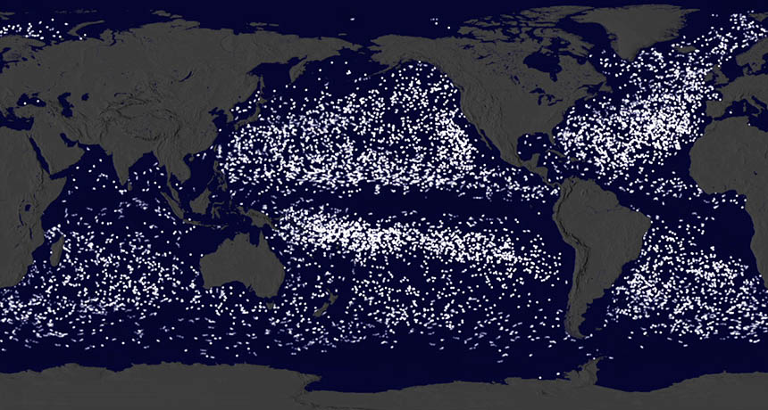
Here’s where 17,000 ocean research buoys ended up

Garbage in, garbage out. But where does all that garbage go? In the oceans, floating bits of debris — everything from plastic bags to Legos — tend to ride along ocean currents to a common destination: one of five major whirling ocean gyres, also known as the ocean garbage patches. Researchers recently got a new look at these gyres thanks to a visualization that combined 35 years’ worth of data on another thing humans drop into the oceans: scientific buoys. The visualization was a finalist in the Data Stories competition sponsored by the American Association for the Advancement of Science. The winners were announced May 5.
Free-floating buoys, released by the National Oceanic and Atmospheric Administration, track temperature, saltiness and other ocean properties. Experts at NASA’s Scientific Visualization Studio combined the movements of more than 17,000 buoys to illustrate the motions of the oceans (see animation below). The buoys start off scattered across the oceans, with some in neat lines that follow the paths of buoy-deploying research vessels. From this chaos, the buoys begin to migrate into clusters. Over time, most drop off the grid and disappear, but some buoys eventually end up in one of the ocean garbage patches.

The garbage patches aren’t floating landfills of intact soda bottles and yogurt cups. The gyres are instead speckled with tiny plastic bits smaller than grains of rice, as many as 100,000 per square kilometer. All that plastic can end up in fish and serves as a foundation for microbe colonies (SN: 2/20/16, p. 20).

Wiping out gut bacteria impairs brain

Obliterating bacteria in the gut may hurt the brain, too.

In mice, a long course of antibiotics that wiped out gut bacteria slowed the birth of new brain cells and impaired memory, scientists write May 19 in Cell Reports. The results reinforce evidence for a powerful connection between bacteria in the gut and the brain (SN: 4/2/16, p. 23).

After seven weeks of drinking water spiked with a cocktail of antibiotics, mice had fewer newborn nerve cells in a part of the hippocampus, a brain structure important for memory. The mice’s ability to remember previously seen objects also suffered.
Further experiments revealed one way bacteria can influence brain cell growth and memory. Injections of immune cells called Ly6Chi monocytes boosted the number of new nerve cells. Themonocytes appear to carry messages from gut to brain, Susanne Wolf of the Max Delbrück Center for Molecular Medicine in Berlin and colleagues found.

Exercise and probiotic treatment with eight types of live bacteria also increased the number of newborn nerve cells and improved memory in mice treated with antibiotics. The results help clarify the toll of prolonged antibiotic treatment, and hint at ways to fight back, the authors write.

Fruit fly’s giant sperm is quite an exaggeration

Forget it, peacocks. Nice try, elk. Sure, sexy feathers and antlers are showy, but the sperm of a fruit fly could be the most over-the-top, exaggerated male ornamentation of all.

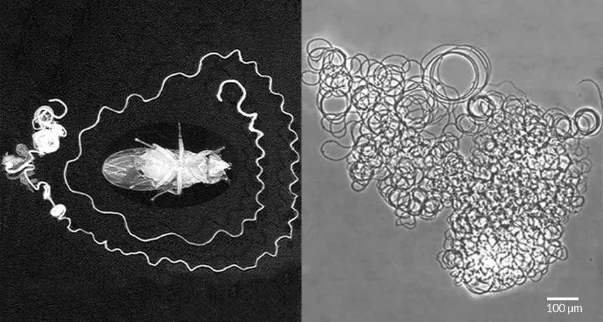
In certain fruit fly species, such as Drosophila bifurca, males measuring just a few millimeters produce sperm with a tail as long as 5.8-centimeters, researchers report May 26 in Nature. Adjusted for body size, the disproportionately supersized sperm outdoes such exuberant body parts as pheasant display feathers, deer antlers, scarab beetle horns and the forward-grasping forceps of earwigs.
Fruit flies’ giant sperm have been challenging to explain, says study coauthor Scott Pitnick of Syracuse University in New York.

Now he and his colleagues propose that a complex interplay of male and female benefits has accelerated sperm length in a runaway-train scenario.

Males with longer sperm deliver fewer sperm, bucking a more-is-better trend. Yet, they still manage to transfer a few dozen to a few hundred per mating. And as newly arrived sperm compete to displace those already waiting in a female’s storage organ, longer is better. Fewer sperm per mating means females tend to mate more often, intensifying the sperm-vs.-sperm competition. Females that have the longest storage organs, which favor the longest sperm, benefit too: Males producing greater numbers of megasperm, the researchers found, tend to be the ones with good genes likely to produce robust offspring. “Sex,” says Pitnick, “is a powerful force.”
Among courtship-oriented body ornaments and weapons (red), the giant sperm of fruit flies (Drosophila) are the most disproportionately exaggerated, according to an index adjusted for body size. Higher numbers (bottom axis) indicate greater exaggeration.

Biologist Kate Rubins’ big dream takes her to the space station

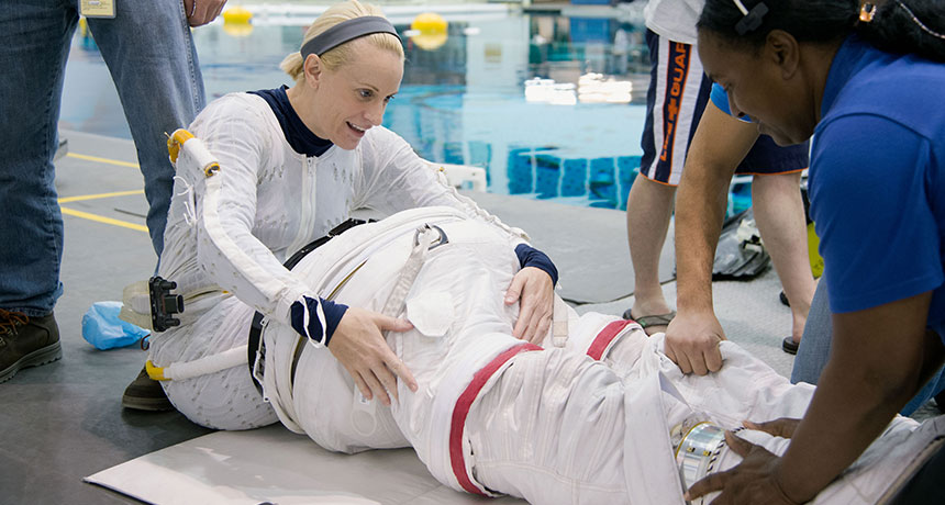
When molecular biologist Kate Rubins blasts off from Kazakhstan on June 24, strapped into the Soyuz spacecraft bound for the International Space Station, the trip will cap off seven years of preparing — and 30 years of hoping.

As a child, Rubins plastered her Napa, Calif., bedroom with pictures of the space shuttle, proudly announcing her intention to be an astronaut. A week at Space Camp in Huntsville, Ala., in seventh grade cemented her vision. But by high school, she concluded that astronaut wasn’t “a realistic job,” she says.
Flash forward to 2009: Rubins is running a lab at the Whitehead Institute for Biomedical Research in Cambridge, Mass., focusing on virus-host interactions and viral genomics. A friend points out a NASA ad seeking astronaut candidates, and Rubins’ long-dormant obsession awakens. Since then, she has learned how to fly a T-38 jet, speak Russian to communicate with her cosmo-naut crewmates, conduct a spacewalk, operate the robotic arm on the ISS and even fix the habitable satellite’s toilet.

Joining NASA meant leaving her 14-person lab behind. But Rubins gained the rare opportunity to collaborate with dozens of scientists in fields as diverse as cell biology and astrophysics. On the space station, she’ll be “their hands, eyes and ears,” conducting about 100 experiments over five months.

She will, for instance, probe how heart cells behave when gravity doesn’t get in the way. And she’ll test a hand-held DNA sequencer, which reads out the genetic information stored in DNA and will be important to future missions looking for signatures of life on Mars.

At times, Rubins will be both experimenter and subject. In one study, she will observe bone cells in a lab dish, comparing their behavior with what happens in a simulated gravity-free environment on the ground. Because astronauts in space are vulnerable to rapid bone loss, CT scanning before and after the mission will also document changes in Rubins’ own hip bone.

Rubins is particularly eager to examine how liquid behaves in microgravity on a molecular scale. In 2013, Canadian astronaut Chris Hadfield created an Internet sensation when he demonstrated that wringing out a wet washcloth in space caused water to form a bubble that enveloped the cloth and his hands. “It’s incredibly bizarre,” Rubins says. Understanding how fluids move in test tubes in space will help NASA plan for Mars exploration, among other applications.
Before any of the research can begin, Rubins has to get off the ground. As treacherous as accelerating to 17,500 miles per hour may sound, she’s not worried.

“An important part of the training experience is making all the information and skills routine,” she says. She predicts that sitting down in the Soyuz spacecraft, pulling out her procedures and getting ready to launch will feel a lot like going into the lab and picking up a pipette — “a normal day at the office.”

Until the engines turn on, anyway. “I think it’s going to feel different when there’s a rocket underneath.”

Francis Crick’s good luck revolutionized biology

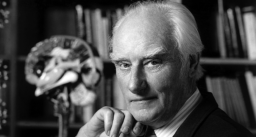
When Francis Crick was 31, he decided he needed to change his luck. As a graduate student in physics during World War II, his research hadn’t gone so well; his experiment was demolished by a bomb. To beat the war, he joined it, working on naval warfare mines for the British Admiralty.

After the war, he sought a new direction.

“There are lots of ways of being unlucky,” Crick told me in an interview in 1998. “One is sticking to things too long. Another is not adventuring at all.”

He decided to adventure.

Molecular biologists everywhere will celebrate that decision on June 8, the centennial of Crick’s birth, in Weston Favell, Northampton, England, in 1916.

“Crick was one of the central figures, one might say the central figure, in the molecular revolution that swept through biology in the latter half of the 20th century,” science historian Robert Olby wrote in a biographical sketch.

In 1953, at the University of Cambridge, Crick and his collaborator James Watson figured out how life’s most important molecule, deoxyribonucleic acid, was put together. DNA, the stuff that genes are made of, became the most famous of biological molecules. Today the image of its double helix structure symbolizes biology itself. It would be easy to make the case that discovering DNA’s structure was the single greatest event in the history of biology — and always will be. In 1962, Watson and Crick won the Nobel Prize for their work (which was, of course, greatly aided by X-ray diffraction imagery from Rosalind Franklin, who unfortunately died before the Nobel was awarded).

Crick’s DNA adventure began at a time when molecular biology was ripe for revolution. But Crick didn’t know that. His choice was lucky.
“I had no idea when I started that molecular biology would advance so fast,” he said. “No idea at all.”

In fact, Crick very nearly chose a different path. His interest in genes was equaled by his curiosity about the brain. Both were topics that he liked to gossip about.

“But I didn’t know enough about either subject,” he said. He just knew a little bit more about biochemistry.

“I thought ‘Well look, I have a training in physics and I know a bit of chemistry, I don’t know anything about the brain.’” So he decided it would be more sensible to start with genes.

“I thought that problem of what genes were and how they replicate and what they did would last me the rest of my life,” he said.

As it happened, genes did occupy him for a couple of decades. Crick made major contributions to elucidating the genetic code during that time. But he never forgot his interest in the brain, and more specifically, consciousness. In the 1970s, he moved from England to California, where he began consciousness research in San Diego at the Salk Institute for Biological Studies.

Consciousness turned out to be a much tougher problem than understanding genes. In retrospect, Crick could see why.

With genetics, “what really made the thing was the simplicity of the double helix. It wrote the whole research program,” he said. “It probably goes back to near the origin of life, when things had to be simple.” Consciousness appeared on the scene only much later, after the evolution of the brain’s vast complexity.

Nevertheless, Crick perceived parallels between genetics and consciousness as subjects for scientific inquiry. As the 20th century came to an end, he mused that consciousness as a concept remained vague — researchers did not all agree about what the word meant. The situation with genes had at one time been similar.

“In a sense people were just as vague about what genes were in the 1920s as they are now about consciousness,” Crick said. “It was exactly the same. The more professional people in the field, which was biochemistry at that time, thought that it was a problem that was too early to tackle. And I think they were right in the ’20s.”

At the end of the 20th century, research on consciousness found itself in much the same state.

“Everybody agrees that it’s an interesting question,” Crick said, “but there are two points of view among scientists: One is that it isn’t a scientific question and is best left to philosophers. And the other one is that, even if it’s a scientific question, it’s too early to tackle it now.”

Crick tackled it anyway. Until his death in 2004, he worked vigorously on the subject with his collaborator Christof Koch, making substantial inroads into identifying the brain activity associated with conscious awareness. Crick was not lucky enough to solve the problem of consciousness, but he perhaps brought the arrival of that solution a little closer.

Ocean plankton held hostage by pirate viruses

When plankton on the high seas catch a cold, the whole ocean may sneeze. Viruses hijacking these microbes could be an important overlooked factor in tracing how living things trap — or in this case, fail to trap — the climate-warming gas carbon dioxide.

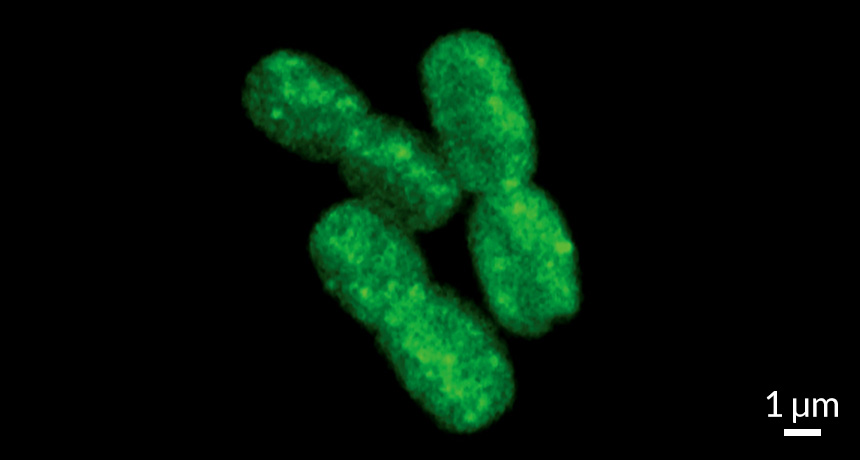
Plants and other organisms that photosynthesize use energy from the sun to capture CO2 for food. The most abundant of these photosynthesizers on the planet are marine cyanobacteria with hardly any name recognition: Synechococcus and Prochlorococcus.
Now, for the first time, a study looks in detail at what happens when some of the abundant viruses found in the sea infect these microbes. Two viruses tested in the lab hijacked cell metabolism, allowing photosynthesis to continue but shunting the captured energy to virus reproduction. The normal use of that energy, capturing CO2, largely shuts down, David Scanlan of the University of Warwick in England and colleagues report online June 9 in Current Biology. As a result, people could be overestimating by 10 percent the amount of CO2 that photosynthesis in the oceans captures.

On any given day, 1 to 60 percent of these plankton may have picked up a viral infection, researchers have estimated. That means that between 0.02 and 5.39 petagrams of carbon — up to 5.39 billion metric tons — may not be captured by marine organisms a year. The high end of that scenario is equivalent to 2.8 times the CO2 routinely captured annually by all the planet’s salt marshes, coral reefs, estuaries, sea grass meadows and seaweeds put together.

Synechococcus and Prochlorococcus plankton “are organisms that you’ve never heard of but you really should have,” says Adam Martiny of the University of California, Irvine. He studies the same kinds of plankton but wasn’t involved in the new virus research, and what he appreciates about it is the intriguing biology of viral manipulation the new work has uncovered.

Until now, Scanlan says, the prevailing view was that while infected plankton were still alive, they were probably carrying on normal photosynthesis. As early as 2003, researchers had clues that the viruses attacking these tiny marine organisms might manipulate photosynthesis in some way, perhaps keeping the process running in an infected cell. These viruses have genes for proteins used in photosynthesis, even though a virus doesn’t even have its own cell much less a way to photosynthesize.

What the viruses are doing, Scanlan and his colleagues have now shown, is subverting their victim’s photosynthesis. Energy capture, the part of photosynthesis directly involved with light, goes on as usual; the cells carry out the routine electron transport for catching energy. But instead of using those sizzling electrons to capture CO2 and turn it into carbohydrates for basic cell metabolism, the viruses shut down this process (called carbon fixation). The light reactions are the ones that researchers normally measure to estimate how much carbon photosynthesis captures in the oceans, but the covert viral shunting means that estimate could be too high.

Scanlan cautions that this is just the beginning of working out the numbers and possible climate effects of virus diseases for these organisms. Whatever the current effects of this takeover turn out to be outside the lab, they may intensify as the climate changes. Synechococcus and Prochlorococcus are “projected to be winners in the new, warmer oceans” and may become even more numerous, Martiny says. And what’s good for them may also increase the abundance of the viral pirates that hijack them.

The 43-year history of journeys to Jupiter, in one graph

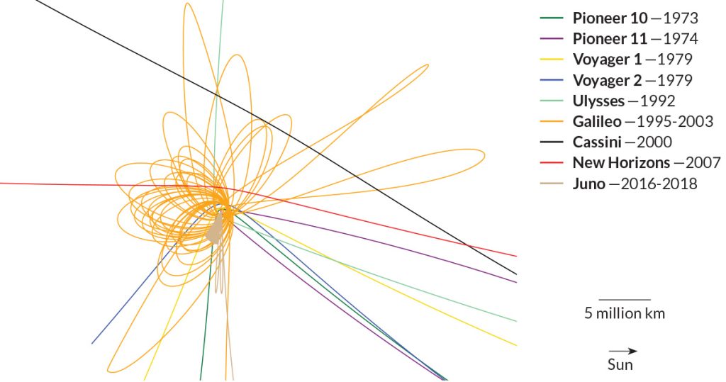
Since 1973, eight spacecraft have flown past or orbited Jupiter. On July 4, NASA’s Juno probe will become the planet’s ninth visitor.

Juno’s trajectory is different than all others, as seen in the plot above and in the video. For 20 months, Juno will repeatedly skim the cloud tops, looping over the poles on orbits that are almost perpendicular to Jupiter’s equator.

Most other spacecraft zipped by, using the planet’s gravity to speed them along to other destinations. Only Galileo, which arrived in 1995, stuck around; it spent nearly eight years circling Jupiter’s equator, repeatedly buzzing the four largest moons.

Baby birds’ brains selectively respond to dads’ songs

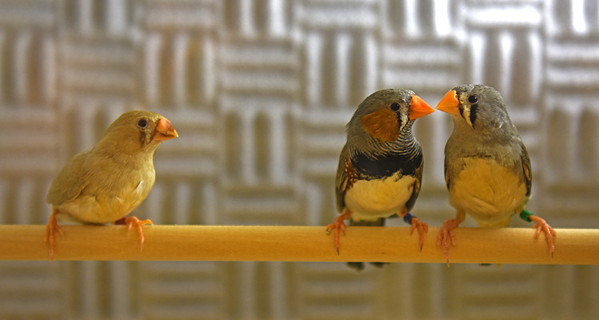
Young zebra finches (Taeniopygia guttata) learn to sing from a teacher, usually dad. Remembering dad’s tunes may even be hardwired into the birds’ brains.

Researchers at the Okinawa Institute of Science and Technology in Japan measured activity in the brains of male juvenile birds listening to recordings of singing adult males, including their fathers. The team focused its efforts on neurons in a part of the brain called the caudomedial nidopallium that’s thought to influence song learning and memory.

A subset of neurons in the caudomedial nidopallium lit up in response to songs performed by dad but not those of strangers, the team reports June 21 in Nature Communications. The more baby birds heard songs, the more their neurons responded and the clearer their own songs became. Sleep and a neurotransmitter called GABA influenced this selectivity.

The researchers suggest that this particular region of the brain stores song memories as finches learn to sing, and GABA may drive the storage of dad’s songs over others.
Researchers played a variety of sounds for young zebra finches: their own song, dad’s song and songs and calls from other adult finches. Over time, their songs became more and more similar to that of their father.

Reptile scales share evolutionary origin with hair, feathers

Hair, scales and feathers arose from one ancestral structure, a new study finds.

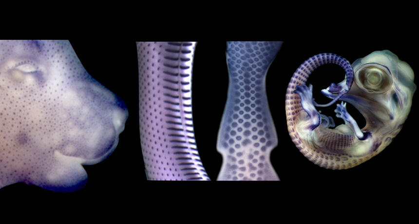
Studies in fetal Nile crocodiles, bearded dragon lizards and corn snakes appear to have settled a long-standing debate on the rise of skin coverings. Special skin bumps long known to direct the development of hair in mammals and feathers in birds also turn out to signal scale growth in reptiles, implying all three structures evolved from a shared ancestor, scientists report online June 24 in Science Advances.
In embryonic birds and mammals, some areas of the skin thicken into raised bumps. Since birds evolved from ancient reptiles, scientists expected that modern snakes, lizards and crocodiles would have the same structures. A study at Yale University last year found that one protein already known to be important in hair and feather development is also active in the skin of developing alligators. But the team did not find the telltale skin thickening. Without that evidence from modern reptiles, scientists weren’t sure if the bumps had been lost in reptiles, or if birds and mammals had evolved them independently, using the same set of genes.
The new results are “a relief,” says Michel Milinkovitch, whose lab led the new study at the University of Geneva. Scientists had come up with a variety of complicated ideas to explain how birds and mammals could share a structure that reptiles lack. But, he says, “the reality is much simpler.”

Clues from a mutant lizard inspired Milinkovitch’s team to probe the mystery. Nicolas Di-Poï, a coauthor of the new study who is now at the University of Helsinki, found that a hair-development gene called EDA was present, but disrupted, in scaleless, or “silky,” bearded dragons. Di-Poï and Milinkovitch searched for similar molecular signals in normal reptile embryos and found genes and proteins associated with hair and feather growth studding the skin. Cell staining revealed characteristic skin thickening at those signal centers.

Reptilian skin bumps eluded previous researchers because they are tiny, appear briefly and don’t all come in at once as they do in mammals, Milinkovitch speculates. “You have to look in the right place at the right time to see them,” he says. “Then boom, you see them, and you’re like, ‘Whoa, they are exactly the same.’”

This study “addresses a fundamental question about identity for skin structures,” says paleontologist Marcelo Sánchez of the University of Zurich, who was not involved in the new research. It’s especially important that the team used crocodiles, lizards and snakes, which are far from typical lab animals, he says. Using nonmodel organisms “gives new insight into evolution we wouldn’t get otherwise.”

The next step is to understand how hairs, feathers and scales diversified from the same ancestral structure. That primordial body covering wasn’t necessarily a scale, says evolutionary biologist Günter Wagner, an author of the 2015 Yale study. “Even though intuitively you would think reptilian-like skin is ancestral, compared to mammals,” he says “it’s entirely unclear what kind of structure the scales and feathers on the one side and hair on the other has evolved from.”